Melanoma
A Guide to Understanding, Identifying, and Detecting the Most Serious Form of Skin Cancer
What Is Melanoma?
Melanoma develops from melanocytes, the pigment‑producing cells that give skin its color. When these cells begin to grow in an abnormal, uncontrolled way—often due to accumulated DNA damage—melanoma can form. Because melanoma has the potential to spread beyond the skin, early detection is critical. It may appear as a brand‑new spot or as a noticeable change in an existing mole, and its color, shape, and size can vary widely.
What Does Melanoma Look Like?
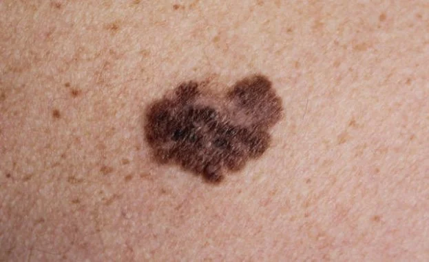
Melanoma can appear in many different ways, which is why it’s often described as the “great imitator” of skin cancer. It may develop as a brand‑new spot or arise within an existing mole that begins to change over time. Because melanoma can vary widely in color, shape, and behavior, recognizing early warning signs is essential.
Melanoma may present as:
A dark or irregularly colored spot
A mole that looks different from your others (“the ugly duckling”)
A lesion with uneven or jagged borders
A mole that contains multiple colors such as brown, black, tan, red, or blue
A pink, red, or skin‑colored growth (amelanotic melanoma), which may be subtle
A spot that grows, darkens, itches, bleeds, or changes in texture
Melanoma can appear anywhere on the body—including areas that receive little sun exposure. On deeper skin tones, melanoma may present as a darker patch, a changing mole, or a new growth on the palms, soles, or under the nails.
Because melanoma does not have a single, predictable appearance, any new, evolving, or unusual spot should be evaluated promptly by a dermatologist.
What Are the Warning Signs of Melanoma?
Dermatologists use the ABCDE criteria to help identify features that may suggest melanoma. These changes can occur in a new lesion or within a mole you’ve had for years.
A — Asymmetry
One half of the lesion looks different from the other. Benign moles are typically symmetrical.
B — Border
Melanomas often have irregular, blurred, or uneven borders. Benign moles usually have smooth, well‑defined edges.
C — Color
Melanomas may contain multiple colors—shades of brown, black, tan, red, white, or blue. Some melanomas have little or no pigment and appear pink or skin‑colored. Benign moles tend to be uniform in color.
D — Diameter
Many melanomas are larger than 6 mm (about the size of a pencil eraser), though they can be smaller. Benign moles are often smaller and more stable.
E — Evolution
Any mole that is changing—growing, darkening, itching, bleeding, or altering in shape—should be evaluated promptly. Benign moles typically remain consistent over time.
The Importance of Ongoing Skin Monitoring
Regular skin checks, sun protection, and self‑monitoring for new or changing lesions are key components of melanoma prevention and early detection. Adults and children with concerning spots, a personal or family history of skin cancer, or increased risk factors can receive thorough evaluation and individualized care.
Expert Evaluation and Treatment of Melanoma in NYC
As a double board‑certified dermatologist and fellowship‑trained Mohs micrographic surgeon, Dr. Adam Nabatian specializes in the evaluation and treatment of melanoma. He performs comprehensive skin examinations, evaluates suspicious lesions, and determines the most appropriate next steps based on the lesion’s features and risk profile.
Schedule Your Melanoma Evaluation in NYC
If you’ve noticed a changing mole, a new spot that looks different from your others, or a lesion that’s evolving over time, a professional skin exam is essential. At Premier Dermatology & Aesthetics, Dr. Adam Nabatian—a double board‑certified dermatologist and fellowship‑trained Mohs micrographic surgeon—provides expert evaluation, diagnosis, and management of melanoma and atypical moles.